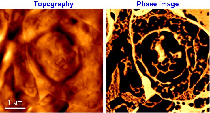
Researchers at Surface Science Western and the Kilee Patchell-Evans Autism Research Group at Western University have developed an atomic force microscope (AFM) phase imaging technique that allows for imaging of subcellular structures in unfixed rat brain sections. The contrast in phase images originates from the difference in mechanical properties between biological structures. Visualization of the native state of biological structures by way of their mechanical properties provides a complementary technique to more traditional imaging techniques such as optical and electron microscopy. The results have been published (PDF) in volume 136 (2011) of the Analyst.

