Electron microscopy can produce stunning images of objects that might look pretty ordinary at first glance. Shea Kraemer, a graduate student from Western University’s Chemistry department, brought SSW some antimony films to image. These films looked dull and black to the naked eye, but when examined in our state-of-the-art instrumentation, the film’s amazing nano-scale structures appeared!
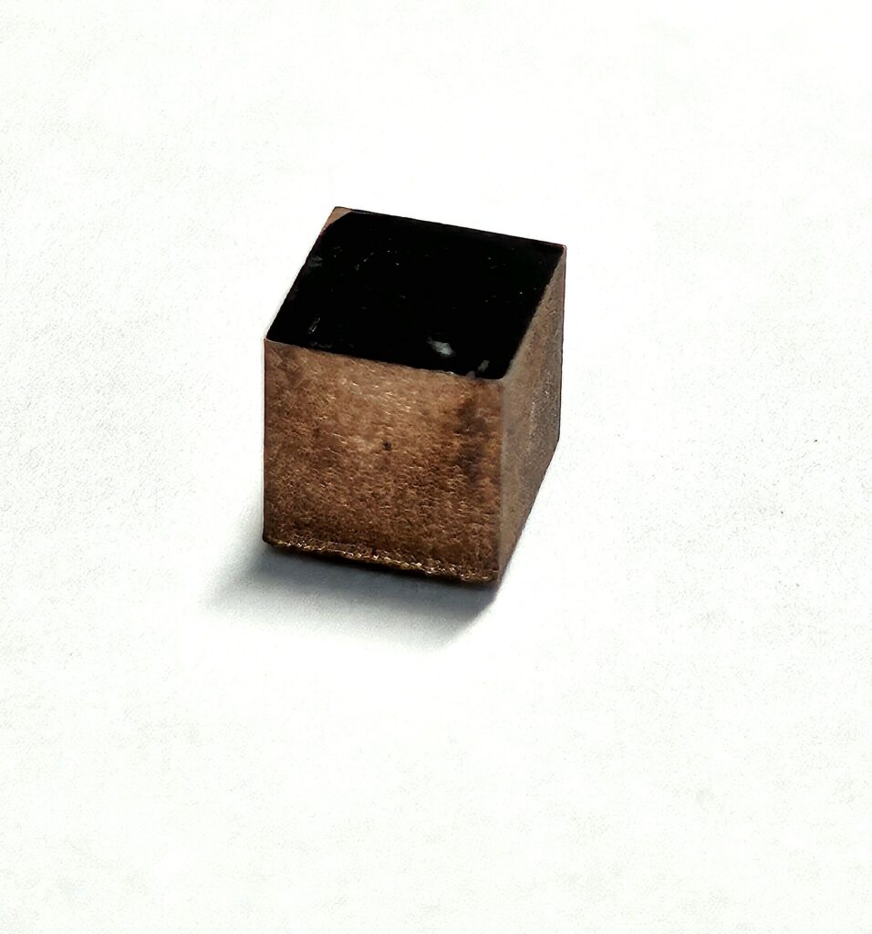
Shea’s current research focuses on enhancing the performance of a potentiometric oxygen/pH sensor using mixed antimony films. A fixed ratio of the antimony (Sb) metal crystals and antimony trioxide (Sb2O3) provide key functional differences to these sensors. Shea required high-resolution imaging to see how the crystals would change through his experiments.
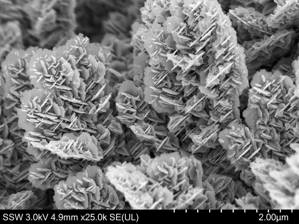
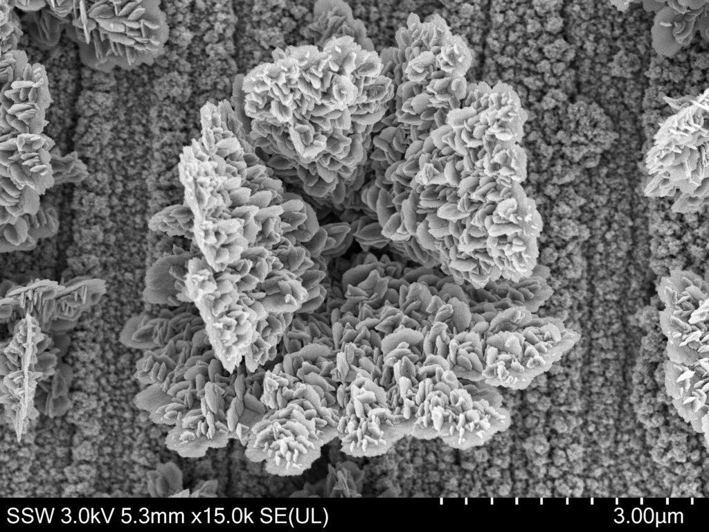
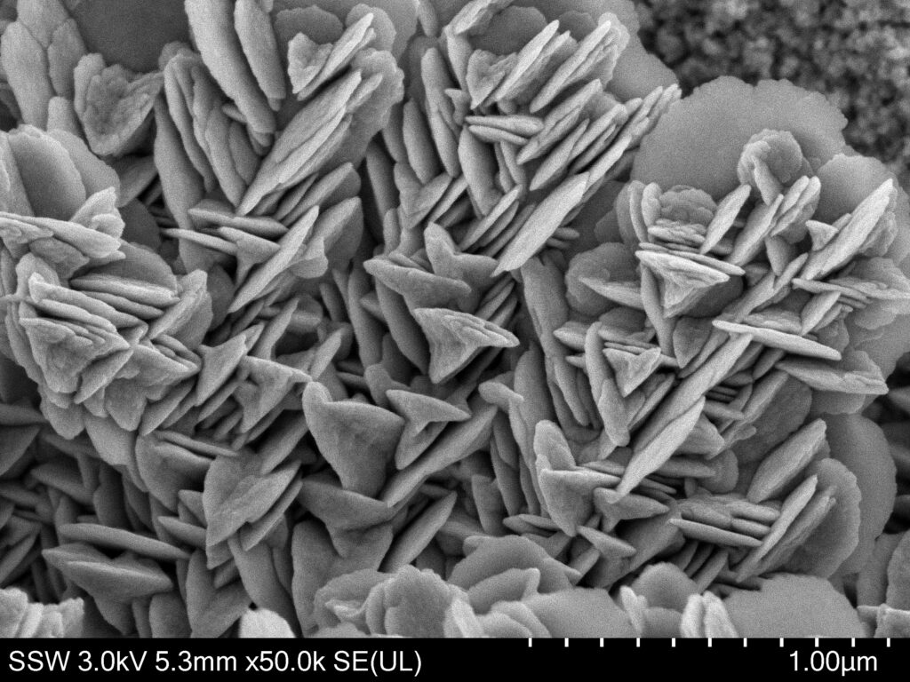
Field emission scanning electron microscopy (FESEM) is an incredible tool that allows the capture of high-resolution/magnification images that have exceptional depth; you can see how the depth of field makes the antimony structures “pop”. These images not only provide detailed insights into the surface morphology, but also bring out the intricate structures of the Sb metal crystals and Sb2O3 film. The depth and clarity of these images will help Shea understand the material’s properties and behaviors better, which is crucial for developing advanced sensors.